

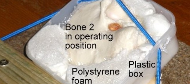
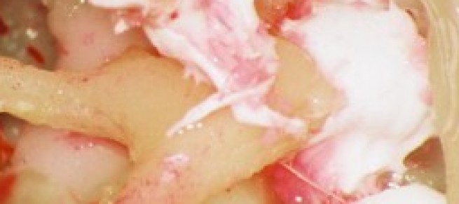
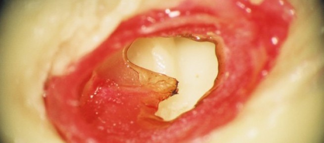
Pettigrew Temporal Bones are plastic castings of cadaver temporal bones and are designed for simulation ear surgery. All surgical operations of the external auditory canal, tympanic membrane, middle ear and mastoid can be practised.
Alastair M Pettigrew is a retired ear surgeon who has 32 years experience in the practical training of ear surgeons and appreciates the difficulties in learning to do ear surgery. He initiated the Glasgow Temporal Bone Course in 1976.
The tiny, complex and variable anatomy of the temporal bone and its diseases provide a challenge for aspiring otologists and a risk for patients. There are textbooks replete with drawings and photographs trying in vain to convey the subtleties of the 3 dimensional anatomy. Practice on cadaver temporal bones is essential but there are difficulties in procurement. An effective way to comprehend the anatomy and surgical operations of the ear is using the temporal bones developed by Mr Pettigrew.
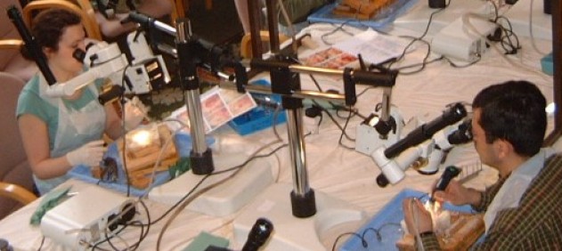
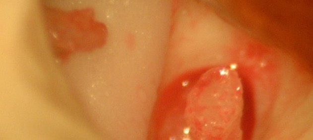
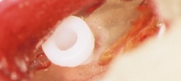
The bones are worked on using a drill, irrigation/suction, operating microscope and ordinary otology instruments. Disease processes such as cholesteatoma, tympanic membrane perforation, ossicular discontinuity and fixed footplate can be incorporated into the bones. It should be noted that there is no internal anatomy of the cochlea or semicircular canals.
The practical skills necessary for ear surgery can be acquired by the aspiring surgeon without putting a patient at risk and greatly reducing the need for practice on cadaver temporal bones.
